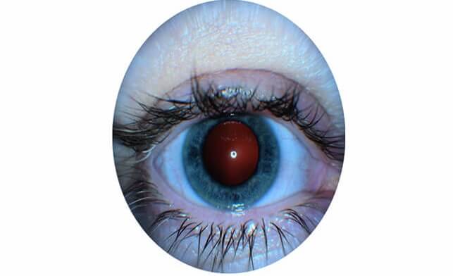
External Eye Imaging
The demand for anterior segment photography is considerably less than for retinal imaging, but this is an important technique for the documentation of anterior segment disease, and should be available in every advanced eye clinic.
Edgbaston Eye Clinic boasts one of the most advanced ocular cameras, the KOWA 3D Camera.
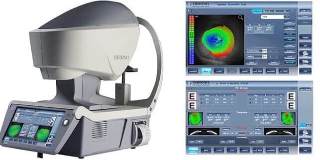
Corneal Topography
Best practice management of keratoconus, including monitoring for disease progression, requires serial corneal tomography assessment. Corneal topography is also used for fitting of lenses, pre-op assessment for laser eye surgery.
At Edgbaston Eye Clinic, we specialise in fitting complex contact lenses and corneal topography is an invaluable tool to enable us to achieve the best contact lens fit. We also use corneal topography to do pre-op laser assessment.
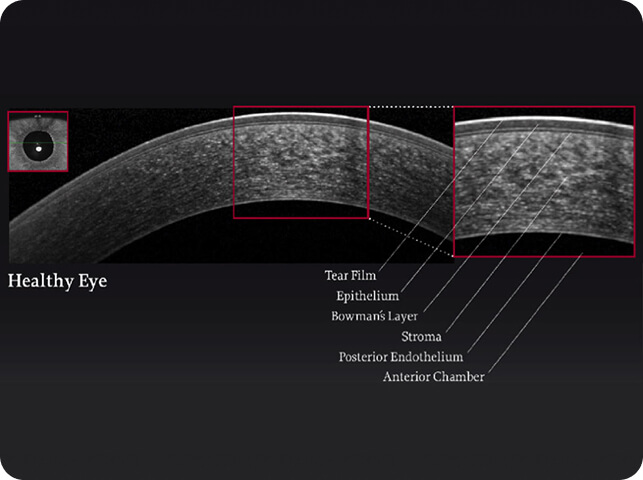
Corneal and Anterior Segment Optical Coherence Tomography (OCT)
OCT uses interferometry to provide high-resolution cross-sectional images of the cornea and anterior segment in a non-invasive manner. Anterior segment OCT is similar to posterior segment OCT although longer wavelength light sources are more commonly used.
Selected Indications and Requirements
The indications for anterior segment OCT are increasing rapidly and access to this modality is recommended for subspecialty eye clinics at tertiary referral centres.
- In glaucoma clinics, anterior segment OCT can be used for direct visualization of the anterior chamber angle, especially in patients with primary angle closure, and also for the diagnosis of patients with plateau iris configuration.
- In corneal and external disease clinics, it can be used to measure peripheral corneal thickness at multiple locations or the depth of anterior stromal corneal scarring useful for planning corneal transplantation surgery.
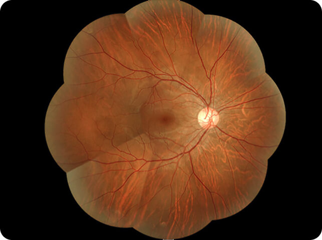
Retinal Imaging
Specially modified cameras may be used to acquire photographs of the ocular fundus.
In combination with light filters and injections of intravenous contrast material, fundus cameras can be used to perform fundus fluorescein angiography (FFA) and indocyanine green angiography (ICG). In combination with light filters alone, they can also be used to perform monochromatic imaging and fundus autofluorescence.
In the UK, a wide range of fundus cameras are available with the KOWA 3D camera as one of the best. At Edgbaston Eye Clinic, patients can see the internal eye in 3D. It is an amazing experience!
The ability to acquire colour photographs of the ocular fundus is an essential part of an advanced eye examination. Colour fundus photography is essential to the diagnosis and monitoring of most posterior segment diseases as well as for disease screening (e.g., as part of the national diabetic retinopathy screening programme in the UK).
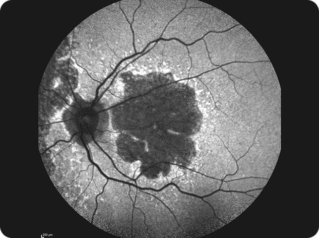
Fundus AutoFluorescence (FAF)
Many structures in the posterior segment possess innate fluorescent properties or “fundus autofluorescence” (FAF), that is, they fluoresce even in the absence of any exogenous contrast agent.
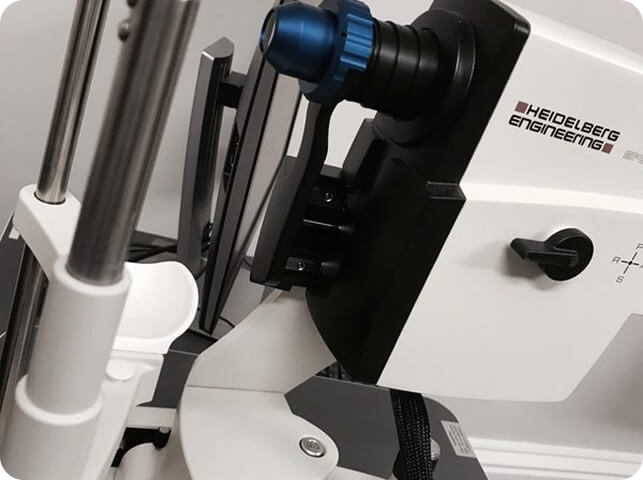
Blue-peak autofluorescence system (Heidelberg Engineering):
- Excitation wavelength of 488nm
- The Spectralis Heidelberg OCT has one of the best autofluorescence modules in all imaging devices
FAF imaging is an essential requirement for highly specialised retinal clinics. It plays a crucial role in the diagnosis and monitoring of patients with inherited retinal disease and in the assessment of patients with toxic retinopathies (e.g., hydroxychloroquine retinopathy).
It also has an emerging role in the diagnosis and monitoring of geographic atrophy in patients with “dry” AMD.
Glaucoma Module Premium Edition (GMPE)
Edgbaston Eye Clinic is proud to be equipped with the Glaucoma Module Premium Edition (GMPE), one of the most advanced glaucoma imaging technologies currently available. Integrated within our state-of-the-art OCT scanner, the GMPE is a highly specialised diagnostic module designed specifically for the early detection, diagnosis and long-term monitoring of glaucoma.
Unlike conventional OCT imaging, the GMPE provides an exceptionally detailed assessment of the optic nerve, retinal nerve fibre layer and macular ganglion cell complex. By combining sophisticated imaging algorithms with advanced analytical software, the system is capable of detecting subtle structural changes that may occur long before a patient notices any visual symptoms.
This technology is particularly valuable in the assessment of patients with ocular hypertension, glaucoma suspects, a family history of glaucoma, and those with established glaucoma requiring long-term monitoring. The enhanced diagnostic precision provided by the GMPE enables more informed clinical decision-making and supports earlier intervention where appropriate.
Our OCT scanner is among the most advanced imaging systems available in clinical practice, and Edgbaston Eye Clinic is the only clinic within the Edgbaston Medical Village equipped with the Glaucoma Module Premium Edition. This investment reflects our commitment to providing the highest standard of specialist glaucoma care using the latest diagnostic technologies.
As glaucoma often develops silently and without symptoms in its early stages, access to advanced imaging technology such as the GMPE plays a crucial role in preserving sight through early detection and ongoing monitoring.