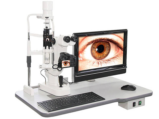
Digital Slit Lamp Examination Birmingham
Detailed microscopic examination of the front and back of the eye at Edgbaston Eye Clinic
A slit lamp is a specialised ophthalmic microscope that provides a highly magnified, three-dimensional view of the structures of the eye. It is one of the most important instruments used during a specialist eye examination.
At Edgbaston Eye Clinic in Birmingham, digital slit-lamp biomicroscopy allows the eye to be examined in considerably greater detail than is possible with the naked eye or a simple handheld instrument.
The examination is painless and normally takes only a few minutes as part of a wider clinical assessment.
What can a slit lamp examine?
The slit lamp produces a narrow, precisely controlled beam of light that can be directed through different structures of the eye. This allows the clinician to assess the:
- eyelids and eyelashes;
- conjunctiva and ocular surface;
- cornea;
- anterior chamber;
- iris and pupil;
- crystalline lens;
- anterior vitreous;
- optic nerve; and
- retina when appropriate specialist viewing lenses are used.
Moorfields similarly describes slit-lamp examination as providing detailed views of structures including the eyelids, cornea, iris, lens and anterior chamber, while specialist lenses allow examination of the back of the eye.
Why is slit-lamp examination important?
Many eye conditions produce subtle physical changes that cannot be identified from symptoms or vision testing alone.
A slit-lamp examination can help identify and assess conditions affecting the front of the eye, including dry eye disease, blepharitis, corneal infections and inflammation, foreign bodies, cataract, uveitis and other anterior eye conditions.
It is also particularly important in glaucoma assessment. Examination of the anterior eye can reveal findings associated with conditions such as pigment dispersion or pseudoexfoliation, while techniques such as Van Herick assessment can help evaluate the depth of the anterior chamber.
Can a slit lamp examine the retina?
Yes.
When combined with specialist diagnostic lenses, slit-lamp biomicroscopy allows a magnified binocular examination of the optic nerve, macula, retina and vitreous.
If a patient presents with symptoms such as new flashes or floaters and a retinal tear is suspected, the College of Optometrists recommends a dilated fundal examination using an indirect viewing technique.
This type of examination can therefore assist in identifying abnormalities including retinal haemorrhage, retinal holes or tears, vitreous changes, optic nerve abnormalities and features of retinal disease.
Importantly, retinal photography and OCT imaging complement rather than replace direct clinical examination. The College notes that fundus imaging does not examine the external eye, anterior chamber or ocular media and therefore does not replace slit-lamp biomicroscopy.
What is a digital slit lamp?
A digital slit lamp combines conventional slit-lamp microscopy with digital imaging.
Where clinically useful, photographs can be recorded to document an abnormality and provide an objective record for future comparison.
This can be particularly helpful when monitoring visible changes involving the:
cornea, ocular surface, eyelids, lens, optic nerve or other observable eye structures.
Digital images can also help when explaining findings to patients or providing supporting clinical information with an onward referral.
Does a slit-lamp examination require dilating drops?
Not always.
Many structures at the front of the eye can be examined without dilation. However, if a detailed examination of the retina, optic nerve or peripheral fundus is required, dilating eye drops may be recommended to provide a more complete view.
The College advises pupil dilation and/or indirect fundus examination when an adequate view of the back of the eye cannot otherwise be obtained.
Digital slit-lamp examination at Edgbaston Eye Clinic
At Edgbaston Eye Clinic, 38 Harborne Road, Edgbaston, Birmingham, B15 3HE, slit-lamp biomicroscopy forms an important part of specialist ophthalmic assessment rather than being used as a standalone test.
Findings can be interpreted alongside other investigations where appropriate, including OCT imaging, Optos ultra-widefield retinal imaging, eye-pressure measurement, gonioscopy and visual-field testing.
The purpose is not simply to obtain an image, but to combine detailed clinical examination with appropriate diagnostic technology to understand what is happening within the eye.
Mr Don Williams ACP (Ophthalmology)
Edgbaston Eye Clinic
The Consulting Rooms
38 Harborne Road
Edgbaston
Birmingham
B15 3HE
Tel: 01217263199