


Birmingham Eye Mole Clinic
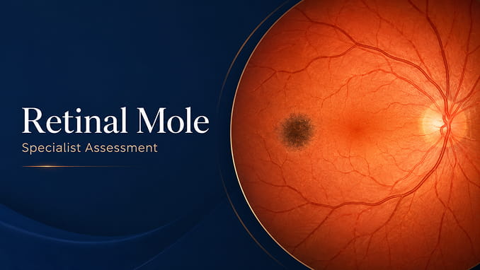
A retinal or eye mole is a common patient description for a pigmented lesion found at the back of the eye. In many cases, the correct clinical term is a choroidal naevus, sometimes described as an eye mole or eye freckle.
Most choroidal naevi are benign and remain harmless throughout life. However, because a small choroidal melanoma can sometimes resemble a naevus, any newly discovered or previously undocumented lesion should be examined carefully and recorded accurately.
At Edgbaston Eye Clinic in Birmingham, retinal mole and choroidal naevus assessments are led by clinical specialist, Mr Don Williams.
The clinic combines specialist dilated retinal examination with Optos ultrawide-field imaging and Heidelberg Spectralis OCT scanning. This allows many lesions to be photographed, assessed and documented so that reassuring features, suspicious features and any future change can be identified more reliably.
Request a Retinal Mole Assessment
What is a retinal mole?
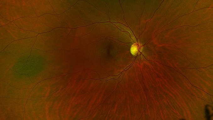
The term “retinal mole” is commonly used to describe a dark or pigmented area seen at the back of the eye.
Many of these lesions are actually located in the choroid, the vascular and pigmented layer beneath the retina. The correct term is therefore usually choroidal naevus.
A choroidal naevus develops from pigment-producing cells called melanocytes. It is similar in principle to a mole or freckle on the skin, although it is located inside the eye and cannot be seen without specialist examination.
Other conditions can also resemble a choroidal naevus. These include variations of the retinal pigment epithelium, congenital hypertrophy of the retinal pigment epithelium, melanocytoma, old haemorrhage and other benign or potentially significant lesions.
This is why the appearance of a dark retinal lesion should be interpreted by an appropriately experienced eye-care clinician rather than judged from a photograph or scan alone.
Is a choroidal naevus dangerous?
Most choroidal naevi are benign and do not threaten sight or general health.
A small minority have unusual or suspicious characteristics that require closer surveillance or onward referral. The purpose of specialist examination is not to suggest that every eye mole is dangerous. It is to distinguish lesions that look typically benign from those that cannot safely be classified without further investigation.
A single assessment may provide substantial reassurance, but comparison with earlier or later retinal images is often particularly valuable. Stability over time is an important feature when assessing many pigmented lesions.
Can an eye mole become melanoma?
Malignant transformation of a typical choroidal naevus is uncommon but possible.
However, small choroidal melanomas and benign naevi can sometimes have overlapping appearances, particularly when no previous retinal photographs are available. The clinically important question is therefore not simply whether a mole can 'turn cancerous but whether the lesion currently has any features associated with increased concern.
These features may include:
- measurable elevation or increasing thickness
- documented enlargement
- orange pigment over the lesion
- fluid beneath or around the retina
- a mushroom-shaped appearance
- associated retinal changes
- new visual symptoms attributable to the lesion
- an appearance that cannot be characterised confidently
The presence of one feature does not automatically mean that melanoma is present. The findings must be considered together and, when necessary, investigated through the appropriate hospital eye or ocular-oncology pathway.
Do retinal moles cause symptoms?
Most choroidal naevi cause no symptoms and are found unexpectedly during an eye examination.
A lesion may occasionally affect vision if it is close to the macula, causes fluid beneath the retina or is associated with another retinal complication.
Possible symptoms requiring assessment include:
- new blurred or distorted vision
- a shadow or missing area in the vision
- new flashing lights
- a sudden increase in floaters
- unexplained reduction in vision
- a change identified during another retinal examination
These symptoms are not specific to choroidal melanoma and are much more commonly caused by other eye conditions. Nevertheless, new symptoms should not be ignored.
The absence of symptoms also does not establish that a lesion is harmless. Some important retinal and choroidal conditions are detected only during a dilated eye examination.
Why has my eye mole only just been discovered?
A choroidal naevus may have been present for many years without being noticed.
It may be discovered for the first time because:
- your pupils have been dilated for a more complete retinal examination
- ultrawide-field imaging has captured an area of the peripheral retina
- a clearer or more comprehensive image has been obtained
- you have attended a different clinic with specialist retinal equipment
- the lesion was not previously photographed or documented
- cataract removal or clearer ocular media have improved the view of the retina
A newly documented lesion is not necessarily a newly developed lesion. When no previous photographs exist, a high-quality baseline image becomes particularly important.
Why does a specialist retinal examination matter?
An eye mole is located inside the eye, so it cannot be inspected or monitored by the patient.
A specialist retinal assessment considers the lesion itself and the surrounding structures. This includes its location, shape, pigmentation, estimated size, margins and relationship to the optic nerve, macula and retinal blood vessels.
The examination also looks for features such as:
- elevation
- subretinal fluid
- orange pigment
- drusen or longstanding retinal pigment changes
- a surrounding halo
- retinal detachment
- haemorrhage or exudation
- changes affecting the overlying retina
- evidence of enlargement when previous images are available
No single scan can reliably answer every clinical question. The findings from retinal examination, photography, OCT and previous records must be considered together.
How is a retinal mole assessed at Edgbaston Eye Clinic?
The assessment begins with a detailed history and specialist examination.
You will be asked whether the lesion has been seen previously, whether earlier photographs are available and whether you have experienced any visual symptoms. Your wider ocular history, medical history and relevant risk factors will also be considered.
The assessment may include the following.
Dilated retinal examination
Dilating drops allow a more complete examination of the retina, choroid, optic nerve and peripheral fundus.
Mr Williams will examine the lesion directly and assess its size, margins, surface appearance, pigmentation and relationship to nearby retinal structures.
Dilation is essential because some lesions cannot be assessed adequately through an undilated pupil.
Optos ultrawide-field retinal imaging
Edgbaston Eye Clinic is equipped with the Optos Daytona ultrawide-field retinal imaging system.
Optos imaging can capture a wide view of the retina in a single image and is particularly valuable for documenting lesions located away from the central retina.
The image can provide a baseline record showing:
- the lesion’s position
- its approximate dimensions
- its relationship to retinal blood vessels and other landmarks
- pigmentation and surface features
- surrounding retinal changes
High-quality baseline photography is one of the most useful tools for detecting enlargement over time. Future images can be aligned and compared rather than relying on memory or written descriptions alone.
Some very anterior lesions may extend beyond the field captured by any retinal camera. Direct clinical examination remains necessary even when ultrawide-field imaging is available.
Heidelberg Spectralis OCT scanning
Optical coherence tomography produces detailed cross-sectional images of the retina.
Where the position of the lesion allows, OCT can help assess:
- the retina overlying the naevus
- subtle subretinal fluid
- disruption or elevation of retinal layers
- retinal pigment epithelial changes
- effects on the macula
- whether the lesion appears flat or elevated within the limits of the scan
OCT is particularly valuable because small amounts of fluid may not always be apparent from colour photography alone.
OCT does not independently diagnose or exclude melanoma. Larger, thicker or very peripheral lesions may require ocular ultrasonography or other investigations within a hospital ophthalmology or ocular-oncology service.
Comparison with previous images
Previous retinal photographs, OCT scans and examination records can be extremely helpful.
A lesion that has remained unchanged over several years is generally more reassuring than one showing definite enlargement. Patients are encouraged to bring or request copies of any previous retinal images where possible.
When earlier images do not exist, the examination at Edgbaston Eye Clinic can provide a detailed baseline for future comparison.
What is the MOLES system?
MOLES is a recognised clinical system or algorithm used to help assess melanocytic choroidal lesions and guide monitoring or referral.
The letters represent:
- M – Mushroom shape
- O – Orange pigment
- L – Large size
- E – Enlargement
- S – Subretinal fluid
Each feature is assessed clinically and through appropriate imaging. The resulting findings help distinguish a common naevus from a lesion requiring closer monitoring, ophthalmological assessment or urgent referral.
MOLES is not a home checklist and should not be used by patients to assess retinal photographs themselves. Image quality, lesion position and clinical interpretation all influence the assessment.
At Edgbaston Eye Clinic, 38 Harborne Road, Edgbaston, Birmingham, B15 3HE recognised risk features are considered alongside the complete retinal examination rather than using any score in isolation.
What happens if the mole appears harmless?
If the lesion has reassuring features, the findings will be explained and an appropriate monitoring plan discussed.
This may include:
- recording a high-quality baseline retinal photograph
- retaining OCT images where the lesion can be scanned
- comparison with previous records
- review at an interval based on the lesion’s features
- advice about symptoms that should prompt an earlier examination
Not every typical naevus requires frequent specialist review. The interval should reflect the appearance of the lesion, whether it has been documented previously and the level of clinical uncertainty.
What happens if the lesion looks suspicious?
A suspicious lesion should not simply be watched indefinitely within a community clinic.
If the lesion has concerning features, appears to have enlarged, cannot be characterised safely or requires investigations not available at Edgbaston Eye Clinic, Mr Williams will explain the findings and arrange referral through the appropriate hospital ophthalmology or ocular-oncology pathway.
The referral can include relevant clinical findings and high-quality retinal or OCT images to help with triage.
Edgbaston Eye Clinic does not replace a specialist ocular-oncology centre. Its role is to provide careful assessment, documentation, risk recognition and timely onward referral when required.
Does every retinal mole need ocular ultrasound?
No. Many flat or minimally elevated choroidal naevi can be assessed and monitored using clinical examination, sequential colour photography and OCT where appropriate.
Ocular ultrasound may be required when:
- the lesion appears significantly elevated
- its thickness cannot be assessed adequately
- OCT cannot image the lesion
- it is very peripheral
- the view is limited
- there are suspicious clinical features
- specialist oncology assessment is required
When ultrasound or specialist tumour assessment is needed, onward referral will be recommended.
Why choose Edgbaston Eye Clinic for retinal mole assessment?
Retinal mole assessment at Edgbaston Eye Clinic is led personally by Mr Don Williams FCOptom, Advanced Clinical Practitioner in Ophthalmology and registered Clinical Specialist.
Mr Don Williams has extensive experience in dilated retinal examination and in identifying pigmented and non-pigmented retinal lesions through hospital and independent practice.
Edgbaston Eye Clinic is particularly well equipped for the initial assessment and documentation of choroidal naevi because it provides:
- specialist dilated retinal examination
- Optos Daytona ultrawide-field retinal imaging
- Heidelberg Spectralis OCT scanning
- detailed baseline documentation
- comparison with previous and future images
- assessment of recognised suspicious features
- clear explanation of findings
- an appropriate onward referral pathway when necessary
The examination is not simply an automated scan. Every image is interpreted in the context of the clinical examination, symptoms, previous records and the appearance of the whole retina.
Patients are seen in an independent private specialist eye clinic without the retail or spectacle-sales environment of a conventional high-street practice.
Eye melanoma in the news
Recent news reports have highlighted patients whose eye melanoma was discovered unexpectedly during an eye examination.
In July 2026, The Independent reported the experience of a 59-year-old man from Stourbridge whose uveal melanoma was detected following a routine eye appointment despite him having no symptoms.
Man who went to opticians for reading glasses diagnosed with melanoma | The Independent
Liverpool University Hospitals has also reported the story of a patient whose ocular melanoma was identified following a routine eye appointment and subsequent hospital assessment.
These cases are unusual and should not lead patients to assume that every eye mole is cancerous. Most choroidal naevi are benign. They do, however, illustrate why examining the health of the retina can be as important as checking the spectacle prescription.
Early recognition of a suspicious lesion allows the patient to enter the appropriate specialist pathway without avoidable delay.
Should I arrange an examination if an optician has found an eye mole?
Yes. A newly discovered or previously undocumented eye mole should be assessed appropriately.
You may particularly benefit from specialist assessment if:
- no retinal photograph was taken
- the lesion has not been measured or documented
- you have not had a dilated retinal examination
- you have been told the lesion is elevated or unusual
- previous images suggest possible change
- you have developed new visual symptoms
- you would like a private second opinion
- you are unsure what follow-up has been recommended
An assessment can often provide reassurance, establish a reliable baseline and identify whether observation or onward referral is appropriate.
Can a retinal photograph diagnose melanoma?
No. A retinal photograph is extremely valuable, but it cannot by itself diagnose or exclude melanoma.
The image must be interpreted alongside a dilated examination, OCT findings where appropriate, previous photographs and the wider clinical picture. Some lesions would require ocular specialist ocular-oncology examination.
The purpose of imaging is to improve detection, documentation and comparison, not to replace clinical judgement.
Book a retinal mole assessment at Birmingham Eye Mole Clinic
If you have been told that you have a retinal mole, eye freckle, pigmented retinal lesion or choroidal naevus, you can request a specialist assessment with Mr Don Williams at Edgbaston Eye Clinic, 38 Harborne Road, Edgbaston, Birmingham, B15 3HE. Tel: 01217263199 or info@edgbastoneyeclinic.com
How much does it cost?
The consultation including all tests required such scans cost £425 payable by card on the day. We advise patients not to drive after the appointment.
To request an appointment, complete the online appointment form or telephone 0121 726 3199.
Request a Retinal Mole Assessment
Clinical guidance and further reading
This page has been informed by current UK guidance and patient information, including:
- Royal College of Ophthalmologists, Referral Pathways for Adult Ocular Tumours
- College of Optometrists, Pigmented Fundus Lesions Clinical Management Guideline
- LOCSU, Naevus Referral Filtering and Monitoring Pathway
- Moorfields Eye Hospital, Essential Adult Ocular Oncology Guide
- NHS information on eye cancer
- Moorfields information on uveal melanoma
Birmingham Eye Mole Clinic at Edgbaston Eye Clinic, located on 38 Harborne Road, Edgbaston, Birmingham, B15 3HE. Tel: 01217263199 or info@edgbastoneyeclinic.com
Reviewed and updated: July 2026