


Wet Macular Degeneration (Wet AMD) — Urgent Assessment in Birmingham
Wet age-related macular degeneration (wet AMD) is a sight-threatening eye condition in which abnormal blood vessels grow beneath the macula, the central part of the retina, and leak blood or fluid. It can develop over days or weeks, and rapid diagnosis is critical because modern treatment can control the abnormal vessels and reduce further sight loss.
Edgbaston Eye Clinic, at 38 Harborne Road, Edgbaston, Birmingham, B15 3HE provides urgent, same-day macular assessment using Spectralis 4D OCT, autofluorescence and multicolour retinal imaging. Assessments are carried out by Mr Don Williams, an Advanced Clinical Practitioner in Ophthalmology and registered specialist with over 20 years' experience, including 13 years in the Hospital Eye Service. A complete macular assessment costs £425, including your consultation and all diagnostic scans. If wet AMD is diagnosed, Mr Don Williams can arrange urgent referral into the retinal service for treatment.
Sudden change in your vision? Do not wait
Please seek urgent eye care today if:
- Your central vision suddenly becomes blurred or distorted
- Straight lines suddenly appear wavy, bent or crooked
- You develop a new dark, grey or missing area in the centre of your vision
- Words or faces appear distorted
- Objects appear smaller or a different shape in one eye
- You notice a sudden difference between the vision in each eye
A sudden deterioration in central vision should never be left until a routine appointment. Contact Edgbaston Eye Clinic promptly on 0121 726 3199 for a same-day urgent assessment.
View the Wet Age Related Macular Degeneration leaflet here
What is wet AMD?
Wet AMD is the less common but more aggressive form of age-related macular degeneration, accounting for around 10–15% of all macular degeneration diagnosed. It develops when abnormal new blood vessels grow beneath the retina at the macula. These vessels are weak and leak blood or fluid, which damages the macula and can cause scarring and rapid loss of central vision.
Unlike dry AMD, which usually progresses slowly, wet AMD can develop very suddenly — sometimes over days. It can now be treated effectively if caught quickly, which is why urgent assessment of any new central vision symptoms is so important.
How does AMD affect vision?
The macula is a small but extremely important area at the centre of the retina, the light-sensing tissue lining the back of the eye. It is responsible for seeing fine detail.
AMD affects only your central vision — the ability to see fine details close up and at a distance. Peripheral (side) vision usually remains normal. Someone with advanced AMD may see the outline of a clock but not be able to tell the time, and may gradually lose the ability to recognise faces. Because peripheral vision is preserved, AMD does not usually cause total blindness, but losing central vision has a major impact on reading, driving and independence which is why early detection matters so much.
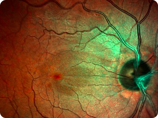
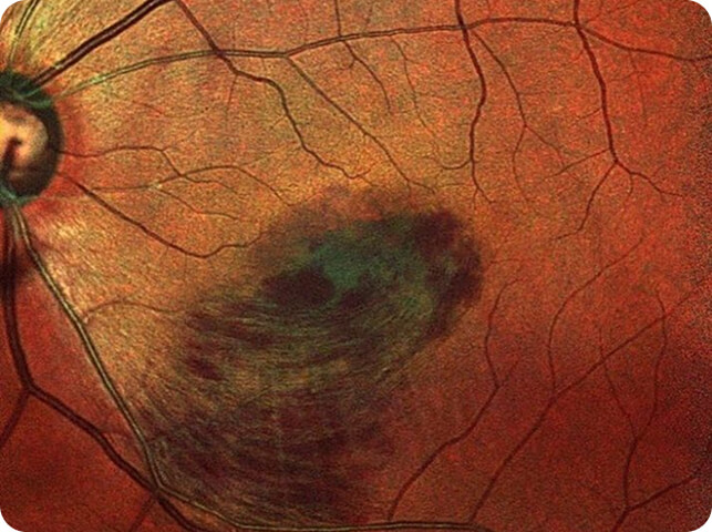
How we diagnose and monitor wet AMD in Birmingham
Edgbaston Eye Clinic uses highly specialised retinal imaging systems to detect the earliest signs of macular change:
- Spectralis 4D OCT scanning — high-density cross-sectional scans of the macula that reveal fluid, bleeding and structural change at the earliest stage
- Autofluorescence imaging — analyses the cellular activity of the retina, helping detect retinal and macular degeneration earlier than standard photography
- Multicolour advanced imaging — highlights different layers of the retina to reveal changes that a single photograph can miss
- Optos or Ultrawide field imaging-captures a view of up to 200° of the retina in a single image, far beyond standard photography, revealing haemorrhages, leakage and the health of the peripheral retina that conventional imaging often miss
If you have been diagnosed with macular degeneration, have your eyes examined and scanned at least once a year — or immediately if you notice any change in your vision. Regular scanning means that if dry AMD ever converts to wet AMD, it is caught at the earliest, most treatable stage.
How is wet AMD treated?
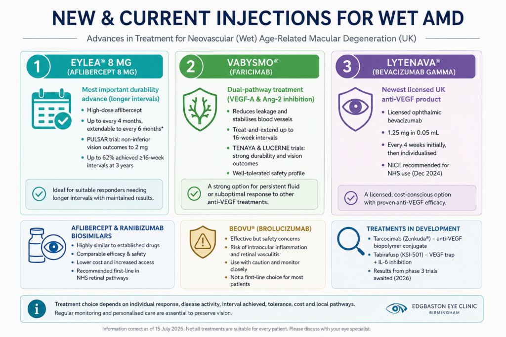
Wet AMD is treated with anti-VEGF injections into the affected eye, which control the abnormal blood vessels and reduce further sight loss. Established drugs include aflibercept (Eylea) and ranibizumab (Lucentis), and newer options — including faricimab, high-dose aflibercept and bevacizumab — have widened the choices available to retinal specialists.
Injection treatment can be provided within private specialist retinal services. The role of Edgbaston Eye Clinic is rapid, accurate diagnosis and urgent referral: if our scans show wet AMD, we refer you into a retinal treatment service without delay, with your imaging included so treatment can begin as quickly as possible. Time matters — the sooner treatment starts, the better the visual outcome tends to be. We can also provide ongoing monitoring alongside your hospital care and continue routine surveillance of your other eye.
How much does a macular assessment cost?
A complete macular assessment at Edgbaston Eye Clinic costs £425. This includes your consultation with Mr Don Williams and all diagnostic imaging — high-density Spectralis OCT scans, autofluorescence and multicolour imaging — with no additional scan fees. You receive a clear explanation of the findings, copies of your report, and, where needed, urgent onward referral for treatment.
Full details of all clinic fees are available on our fees page.
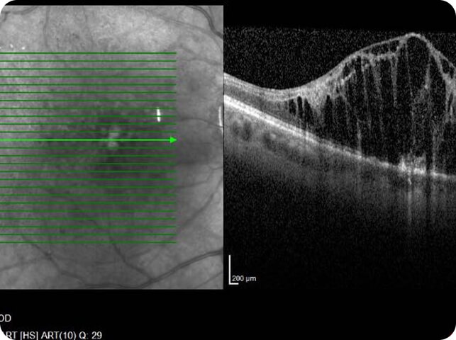
Conditions that can mimic wet AMD
Not every sudden distortion or blur in central vision is wet AMD. Several other macular conditions produce very similar symptoms, and telling them apart matters — because they have different causes, different treatments, and very different levels of urgency:
Central serous retinopathy (CSR) — fluid collects under the macula, typically in younger adults, causing central blur or distortion that can look strikingly like wet AMD on symptoms alone. Unlike wet AMD, CSR often resolves on its own, and the management pathway is entirely different — so distinguishing the two prevents unnecessary alarm and unnecessary treatment.
Retinal vein occlusion with macular oedema — a blocked vein causes swelling and haemorrhage at the macula, blurring central vision.
Diabetic macular oedema — fluid at the macula from diabetic changes, producing gradual or sudden central blur in people with diabetes.
Myopic macular changes — in very short-sighted eyes, abnormal vessels or stretching at the macula can mimic AMD in a younger age group.
Epiretinal membrane, vitreomacular traction and macular hole — mechanical changes at the surface of the macula that distort vision, causing the same wavy lines and reading difficulty patients associate with AMD.
Pattern dystrophies and adult vitelliform changes — inherited or age-related deposits at the macula that can closely resemble AMD on basic examination.
High-resolution OCT, autofluorescence, multicolour and ultra-widefield imaging usually distinguish these conditions clearly — which is why a proper macular assessment, interpreted by an experienced clinician, matters more than any single symptom. Getting the diagnosis right first time means the right pathway from the start: urgent referral for injection treatment where it is genuinely wet AMD, and reassurance, monitoring or a different referral route where it is not.
Medical retina expertise at Edgbaston Eye Clinic
Macular assessment at the clinic is carried out by Mr Don Williams, who also holds Professional Postgraduate Qualification in Medical Retina with Distinction and worked in hospital medical retina services. That experience — seeing wet AMD, CSR and their mimics side by side in a treatment setting — is what informs the clinic's diagnostic approach today: careful imaging, careful interpretation, and an honest answer about what your symptoms are and are not, with urgent referral into treatment on the same day where it is needed.