Everyone will develop presbyopia by the fifth decade of life; however, the age of onset for the signs and symptoms of presbyopia varies between individuals. Refractive error, preferred near working distance, an individual’s stature and ambient lighting all impact the timing of presbyopic signs and symptoms.
Presbyopia is the loss of the ability to read at a normal working distance when fully corrected for distance vision. Presbyopia affects 100% of the population by the fifth decade of life. The anatomy and optics of the eye are crucial for understanding the basis for presbyopia and the onset of its related signs and symptoms.
Anatomy of the eye
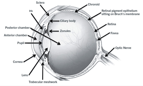
A cross section of the eye is shown in Fig. 1. The eye has an axial length of approximately 23 mm. The clear front part of the eye is the cornea. Similar to the glass of a watch, it is transparent which allows light to travel into the eye. The cornea, with a central thickness of approximately 550 microns, consists of an outer epithelial layer, a middle collagen layer and an inner endothelial layer. The epithelial layer, a barrier that prevents water from entering the cornea, is constantly being replaced every 7 to 10 days. The middle layer consists of collagen fibers that are uniformly arranged making the cornea transparent. In contrast to the cornea, the sclera, the white part of the eye, appears white because light is scattered from its unorganized cross-linked collagen fibers. On the inner surface of the cornea there is a single layer of endothelial cells. These cells do not regenerate and the number of endothelial cells slowly declines with age. The function of the endothelial cells is to pump water out of the cornea. If the endothelial cells are damaged then the cornea swells and becomes cloudy causing a marked decrease in visual acuity.
Located behind the cornea are the anterior chamber, iris, lens, posterior chamber and retina. The anterior chamber is filled with a clear fluid, aqueous humor, containing salts and amino acids to supply nutrients to the cornea and lens. The aqueous humor is constantly being produced by the ciliary body and drained from the eye through the trabecular meshwork. If drainage of the aqueous humor is blocked, the pressure within the eye increases which can result in glaucoma. The iris, the colored part of the eye controls the size of the pupil, constricting in bright light and dilating in dim light. Suspended behind the iris is the crystalline lens. The lens, a transparent, encapsulated biconvex spheroid, consists totally of epithelial cells; however, unlike the epithelium of skin, which sheds, the lens is encapsulated so it continues to grow throughout life. At birth the lens has an equatorial diameter of approximately 6 mm that increases in the adult to approximately 9 mm.
Read more here: http://pointsdevue.com/article/early-signs-and-symptoms-presbyopia