Glaucoma Investigation
Protect your sight with advanced Glaucoma Investigation – specialist testing in Birmingham
- Glaucoma is a potentially a blinding condition
- Primary Open Angle Glaucoma is the second most common cause of irreversible blindness in the UK, the first being Age-Related Macular Degeneration
- Responsible for 12 out every 100,000 people aged 40 and over are certified as sight impaired or severely sight impaired from POAG
- Painless condition and patients may be unaware that they glaucoma until they have started to lose vision
Glaucoma risk factors include:
- High eye pressure
- Increasing age
- Family history
- Shortsighted prescription (>-4)
- Diabetes
- High blood pressure
- Thin cornea
- Afro-Caribbean ancestry
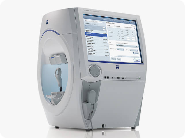
Humphrey Visual Field Analyser 3
- The visual field test measures how far you can see in your peripheral vision (side vision), which is often affected by glaucoma
- Glaucoma typically reduces your peripheral vision first, making it harder to notice the loss until significant damage has occurred
- In advanced stages of glaucoma, the loss of peripheral vision can become so severe that it may feel like looking through a ‘loo roll’ or a narrow tunnel
- Patients often do not notice vision loss until the damage has progressed significantly, making regular visual field testing critical for early detection
- The visual field test can also detect other neurological conditions, such as brain tumours, and is crucial for patients with sudden headaches or visual migraines
- At Edgbaston Eye Clinic, we use the latest Visual Field Analyser technology to provide accurate and comprehensive testing for glaucoma and other conditions
- Visual field testing is painless and plays a vital role in diagnosing glaucoma early to prevent permanent vision loss
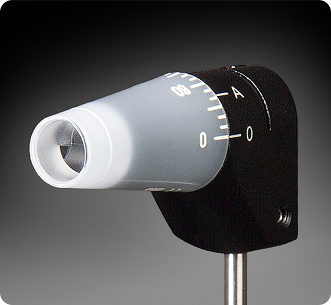
Goldmann Applanation Pressure
- Eye pressure, also known as intraocular pressure (IOP), is the fluid pressure inside the eye, and maintaining a healthy level is crucial for eye health
- High intraocular pressure (IOP) is one of the most significant risk factors for developing glaucoma, especially open-angle glaucoma, the most common form
- Normal eye pressure typically ranges from 10 to 21 mmHg, but individuals can still develop glaucoma even if their eye pressure is within this range
- Elevated IOP occurs when the fluid inside the eye, called aqueous humour, does not drain properly, leading to pressure build-up and potential damage to the optic nerve
- Ocular hypertension is a condition where eye pressure is higher than normal but there are no signs of optic nerve damage or vision loss, which still requires monitoring to prevent glaucoma
- Regular eye pressure checks are essential, especially for individuals over 40 or those with risk factors like family history, African-Caribbean ethnicity, or diabetes
- Goldmann Applanation Tonometry (GAT) measures the force required to flatten a small part of the cornea, making it the most reliable and precise method for measuring IOP
- Routine IOP monitoring is critical because early detection of increased eye pressure can prevent permanent vision damage caused by glaucoma
- Other tonometry methods, such as non-contact (air puff) tonometry, are useful for screening but are less accurate than Goldmann Applanation Tonometry
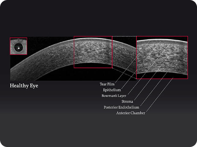
Corneal thickness
- Central Corneal Thickness (CCT) plays a crucial role in determining the accuracy of intraocular pressure (IOP) measurements, which is vital for diagnosing glaucoma
- A thicker cornea can cause artificially high pressure readings, while a thinner cornea may lead to lower pressure readings, potentially masking glaucoma risk
- Measuring the thickness of the cornea is an essential part of any thorough glaucoma investigation, as it provides context for IOP readings and helps determine your true glaucoma risk
- At Edgbaston Eye Clinic, we utilise advanced ophthalmic diagnostic equipment to precisely measure central corneal thickness, ensuring accurate and reliable glaucoma assessments
- CCT measurement is painless and helps create a comprehensive picture of your eye health, allowing for better-tailored glaucoma management and treatment options
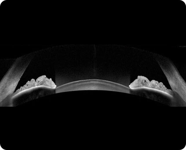
Gonioscopy-analysis of the drainage system of the eye
- At Edgbaston Eye Clinic, we use advanced technology to scan and visualize the drainage system of the eye, allowing for a thorough assessment of the anatomical structures responsible for fluid drainage
- Gonioscopy is an essential test in the evaluation of glaucoma, as it helps assess the functionality of the drainage angle, where fluid leaves the eye
- This test involves gently placing a specialised lens on the eye after numbing the cornea. The lens, cushioned with tear gel, provides comfortable and painless imaging for the patient
- We are fortunate to offer video gonioscopy, allowing us to capture real-time video of the drainage angle in action, enhancing the accuracy of our assessments
- Gonioscopy is crucial for identifying conditions like angle-closure glaucoma, where the drainage system becomes blocked, leading to increased eye pressure
- Our clinic’s state-of-the-art diagnostic equipment ensures a precise evaluation of your eye’s drainage system, helping to provide tailored treatment options for glaucoma

Retinal Nerve Fibre Layer (RNFL) analysis
- The Retinal Nerve Fibre Layer (RNFL) is highly susceptible to damage caused by glaucoma, and early detection is crucial to prevent vision loss
- Conventional tests may not detect RNFL damage until approximately 30% of the nerve layer is already damaged, making early detection difficult
- At Edgbaston Eye Clinic, we use advanced RNFL scanning technology to detect damage at the earliest stage, allowing for prompt intervention
- RNFL scans provide a detailed view of the nerve fibre layer inside the eye, enabling us to identify glaucoma-related damage well before it becomes noticeable through conventional testing
- Early detection of RNFL damage means glaucoma treatment can begin sooner, improving the likelihood of preserving vision and preventing further deterioration
- Significant damage to the nerve fibre layer is irreversible and can be sight-threatening, highlighting the importance of regular RNFL testing, especially for high-risk individuals
- Glaucoma management is far more effective when diagnosed early, and the RNFL scan plays a vital role in safeguarding your vision
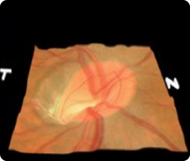
Optic Nerve Imaging
- KOWA stereo imaging system
- At Edgbaston Eye Clinic, we are the only private clinic in Edgbaston offering the advanced Optos (Daytona) ultrawide field imaging system for both retinal imaging and optic disc capture, crucial in the early detection and management of glaucoma
- The Optos (Daytona) provides a 200-degree view of the retina, enabling comprehensive evaluation of the optic nerve head and retinal nerve fibre layer (RNFL), areas commonly affected by glaucoma
- This imaging system allows us to detect optic disc changes, such as cupping, which is a key indicator of glaucoma progression
- The high-resolution images captured by the Optos help us identify and monitor conditions like glaucoma, diabetic retinopathy, and macular degeneration, ensuring accurate diagnosis and treatment
- Optos imaging is non-invasive, fast, and provides immediate results, making it an essential tool for both routine eye exams and detailed glaucoma assessments
- By utilising this cutting-edge technology, Edgbaston Eye Clinic offers unparalleled glaucoma care and optic disc monitoring, ensuring the best outcomes for our patients