Medical Retina
Management of conditions of the back of the eye using eye drops or lasers
- Diabetic eye disease
- Wet age related macular degeneration
- Blockage of blood vessels inside the eye
- Inflammation of the back of the eye
Pupil Dilation Procedure
We often put drops to open the pupil to get a better view of the internal structures of the eye.
It takes about 15 minutes for the drops to work and approximately 2–4 hours to wear off.
We advise all of our patients NOT to drive to the appointment, as the drops will make driving vision very difficult.
Blue Laser Autofluorescence
BluePeak is a non-invasive, scanning laser fundus imaging modality that reveals metabolic stress in the retina using lipofuscin as an indicator.
It offers diagnostic insights into retinal conditions such as Age Related Macular Degeneration and other hereditary diseases affecting the retina.
Video of progression of AMD using Blue Peak Autofluorescence

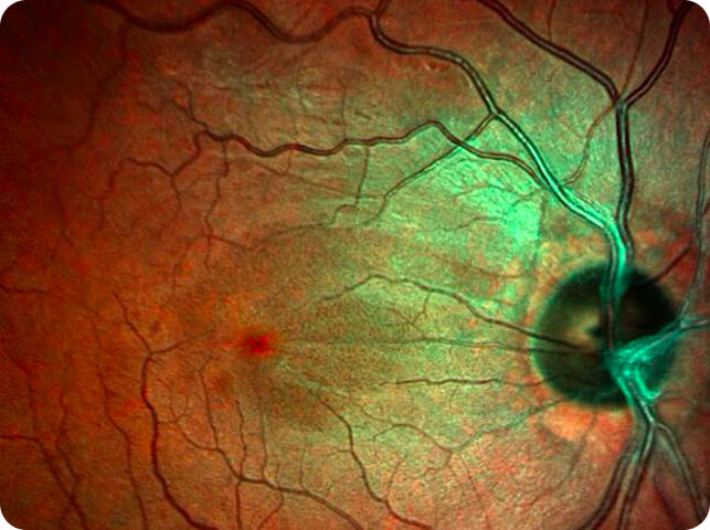
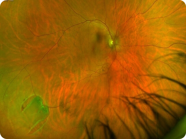
Multicolour Fundus Imaging
Uses three laser wavelengths simultaneously to provide diagnostic images that show distinct structures at different depths within the retina.
The high-resolution, detailed MultiColour images can highlight structures and pathologies not visible with a standard camera.
Ultra Wide Field Imaging
Provides a broad view of the retina to detect peripheral retinal conditions that may be missed with conventional imaging.
