


Optos Ultra-Widefield Retinal Imaging in Birmingham
Edgbaston Eye Clinic is equipped with Optos ultra-widefield retinal imaging, allowing up to approximately 200° of the retina to be captured in a single, non-contact image. This provides a far wider view than conventional retinal photography and is particularly valuable when assessing pathology located away from the central retina.
Ultra-widefield imaging allows us to document, compare and monitor retinal findings over time and forms an important part of the assessment of a wide range of retinal and ocular conditions.
Why Optos imaging is useful
Many important retinal abnormalities occur in the peripheral retina and may not be adequately documented using standard fundus photography alone. Optos imaging can assist with the detection and monitoring of conditions including retinal tears, peripheral retinal degeneration, diabetic retinal changes, vascular abnormalities, inflammatory retinal disease, pigmented retinal lesions such as eye moles or choroidal naevi.
Because the images are digitally stored, they can also provide an invaluable baseline record. This allows future images to be compared carefully for any meaningful change in size, appearance or morphology.
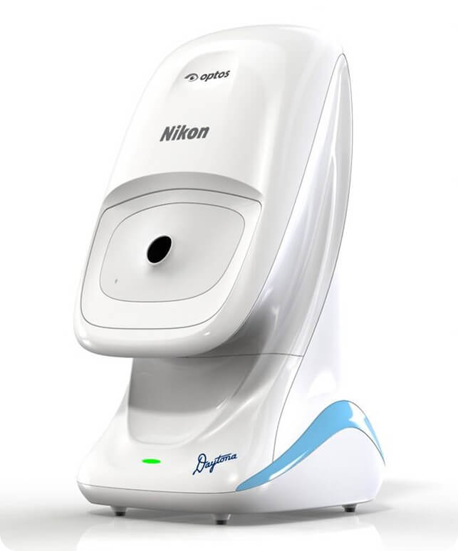
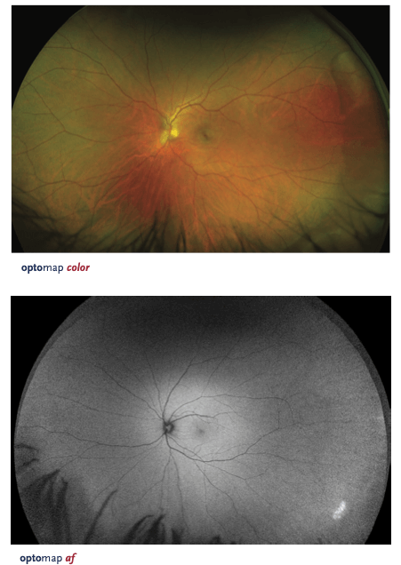
Optos Imaging for Eye Moles and Choroidal Naevi
At the Birmingham Eye Mole Clinic at Edgbaston Eye Clinic, Optos ultra-widefield imaging is used as part of the assessment and surveillance of eye moles, choroidal naevi and other pigmented ocular lesions.
Many pigmented lesions are located in the mid-peripheral or far-peripheral fundus, where ultra-widefield imaging can be particularly useful. Detailed photographic documentation allows the lesion's location, pigmentation, borders and overall appearance to be recorded and compared at subsequent examinations.
Where clinically appropriate, Optos imaging is combined with dilated retinal examination, OCT imaging and structured clinical risk assessment, including assessment for suspicious features such as growth, orange pigment, subretinal fluid or abnormal morphology.
Most eye moles are benign, but accurate documentation and appropriate surveillance are important in identifying the small number of lesions that require closer observation or onward specialist assessment.
More Than a Retinal Photograph
Optos imaging is not simply a photographic record. It forms part of a wider clinical assessment and must be interpreted alongside the patient's symptoms, ocular history, dilated retinal examination and other investigations where appropriate.
At Edgbaston Eye Clinic, ultra-widefield retinal imaging may be used in patients presenting with:
- flashes and floaters
- suspected retinal tears or peripheral retinal degeneration
- diabetic retinal disease
- retinal vascular abnormalities
- inflammatory retinal conditions
- unexplained visual symptoms
- pigmented retinal lesions
- eye moles and choroidal naevi
- medical retina concerns requiring detailed documentation
Fundus Autofluorescence
Our Optos system also provides fundus autofluorescence imaging, which can help demonstrate changes within the retinal pigment epithelium and may provide additional information in selected retinal and macular conditions.
Autofluorescence can be useful in the assessment of conditions affecting retinal pigment metabolism and can complement conventional retinal imaging and OCT where clinically indicated.
Specialist Retinal Assessment in Birmingham
Optos imaging is available as part of a specialist retinal or comprehensive ophthalmic assessment at Edgbaston Eye Clinic, 38 Harborne Road, Edgbaston, Birmingham, B15 3HE.
Where a retinal lesion or pigmented ocular lesion requires closer monitoring, serial imaging can be used to establish a high-quality baseline and assess for future change.