


Optical Coherence Tomography (OCT)
Advanced retinal, macular and optic nerve imaging at Edgbaston Eye Clinic
Optical Coherence Tomography, usually known as OCT, is a non-invasive imaging technique that produces highly detailed cross-sectional images of the structures inside the eye.
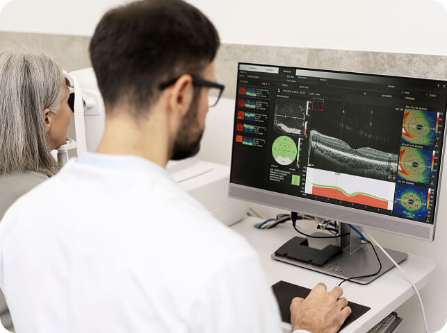
At Edgbaston Eye Clinic in Birmingham, we use the Heidelberg SPECTRALIS OCT, an advanced ophthalmic imaging system capable of producing high-resolution images of the individual layers of the retina and optic nerve. Heidelberg SPECTRALIS technology can visualise multiple retinal layers and is designed to help clinicians identify and monitor subtle structural changes over time.
What does an OCT scan show?
The retina is the light-sensitive tissue at the back of the eye. While conventional retinal photography provides an image of its surface appearance, OCT allows us to examine the retina in cross-section, providing detailed information about its individual layers.
OCT uses light waves rather than X-rays to create these images and is quick and non-invasive. NHS ophthalmology services similarly describe OCT as providing high-resolution cross-sectional images that help clinicians evaluate the eye and make management decisions.
An OCT examination can provide detailed assessment of structures including:
- the macula, responsible for detailed central vision;
- the retinal nerve fibre layer (RNFL);
- the ganglion cell complex;
- the optic nerve head;
- the individual layers of the retina; and
- the retinal structures surrounding certain lesions or areas of abnormality.

What eye conditions can OCT help assess?
OCT is particularly valuable in the investigation and monitoring of many retinal and optic nerve conditions, including:
Glaucoma and ocular hypertension
OCT can provide objective measurements of the optic nerve, retinal nerve fibre layer and ganglion cells. It is an important part of modern glaucoma assessment when interpreted alongside eye pressure, visual fields, optic nerve examination and other clinical findings. NICE includes OCT or optic nerve imaging within glaucoma investigation where available.
Age-related macular degeneration (AMD)
OCT can identify structural changes affecting the macula and detect fluid within or beneath the retina.
Diabetic eye disease
OCT is particularly useful for assessing the macula and identifying retinal thickening or fluid associated with diabetic macular disease.
Epiretinal membranes and macular holes
Cross-sectional imaging can demonstrate changes at the vitreoretinal interface that may not be fully appreciated from retinal photography alone.
Retinal vascular disease
OCT can help identify and monitor macular swelling and other retinal changes associated with conditions such as retinal vein occlusion.
Choroidal naevi and pigmented lesions
Where the position of a retinal or choroidal lesion allows, OCT can provide useful information about the overlying retina and identify features such as subretinal fluid. OCT forms part of the multimodal assessment available through our Birmingham Eye Mole Clinic, but it does not diagnose or exclude melanoma on its own.
Why is OCT useful for monitoring?
One of the major advantages of OCT is the ability to compare scans over time.
A high-quality baseline scan provides an objective record of the retina or optic nerve. Subsequent scans can then be compared with earlier examinations to help identify genuine structural change.
This can be particularly valuable in chronic conditions such as glaucoma, where small changes may develop gradually. Heidelberg specifically highlights the ability of SPECTRALIS imaging to detect and follow subtle structural changes.
Does an OCT scan replace an eye examination?
No.
OCT is an extremely valuable diagnostic tool, but an OCT scan should never be interpreted in isolation.
The scan needs to be considered alongside the patient's symptoms, clinical history, visual acuity, retinal or optic nerve examination and any other appropriate investigations.
For example, glaucoma cannot be diagnosed simply from an OCT result. Clinical optic nerve assessment, visual-field testing and intraocular-pressure measurement remain important components of a comprehensive glaucoma investigation.
This is why the quality of the clinical interpretation is as important as the quality of the equipment.
Is OCT safe?
Yes. OCT is non-invasive and uses light waves to produce images. It does not use X-rays or ionising radiation.
The scan itself usually takes only a short time and does not touch the eye.
Depending on the reason for your examination, dilating eye drops may still be required as part of the wider clinical assessment.
Heidelberg SPECTRALIS OCT at Edgbaston Eye Clinic
At Edgbaston Eye Clinic, OCT imaging forms part of our advanced diagnostic assessment for conditions including glaucoma, macular disease, medical retina and selected choroidal or retinal lesions.
Our Heidelberg SPECTRALIS system is used alongside other specialist diagnostic technology, including Optos ultra-widefield retinal imaging, Humphrey visual-field analysis and detailed clinical examination.
OCT examinations and clinical interpretation are undertaken as part of specialist eye assessment led by Mr Don Williams FCOptom, Advanced Clinical Practitioner in Ophthalmology.
Edgbaston Eye Clinic is located on 38 Harborne Road, Edgbaston
Birmingham
B15 3HE