


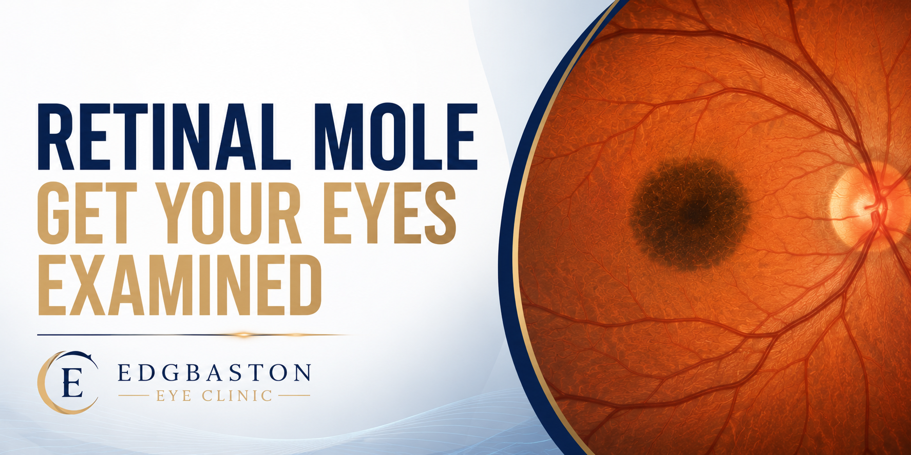
A recent news story highlighted the importance of routine eye examinations after a man attended for what he thought was a simple sight test, only to discover he had a retinal mole, also known as a retinal naevus. Further specialist assessment led to the diagnosis of uveal melanoma, a rare form of eye cancer.
Man who went to opticians for reading glasses diagnosed with melanoma | The Independent
Although this type of cancer is uncommon, the story serves as an important reminder that a comprehensive retinal examination can sometimes detect serious eye conditions before any symptoms develop.
What is a retinal mole?
A retinal mole, more accurately known as a choroidal naevus, is a pigmented lesion found beneath the retina within the choroid, the vascular layer at the back of the eye.
These lesions are relatively common and, in the overwhelming majority of cases, are completely benign. Many people are unaware they have one because they usually cause no pain, no change in vision and no other symptoms.
Most retinal moles never require treatment. However, they should be examined carefully and, in some cases, monitored over time.
Can a retinal mole become cancerous?
Only a small proportion of retinal moles develop into uveal melanoma, the most common primary cancer arising inside the eye in adults.
Although the risk is low, recognising suspicious features early is extremely important because prompt referral to an ocular oncology specialist allows appropriate monitoring or treatment if required.
Specialists assess retinal moles for recognised risk factors such as:
- Thickness of the lesion
- Subretinal fluid
- Orange pigment
- Symptoms
- Growth over time
- Ultrasound characteristics
- Proximity to the optic nerve
These clinical features help determine whether a lesion is likely to be a harmless naevus or requires closer investigation.
Why are retinal examinations so important?
Many retinal conditions develop silently.
A retinal mole, retinal tear, diabetic eye disease or even early ocular melanoma may cause no symptoms whatsoever.
This is why a detailed retinal examination is about much more than checking whether you need new glasses.
Modern retinal assessment allows eye care professionals to examine the health of the retina, optic nerve and macula in remarkable detail, often identifying conditions before they begin to affect vision.
Advanced retinal imaging
At Edgbaston Eye Clinic, retinal examinations are supported by advanced ophthalmic imaging, including:
- Optos Daytona ultra-widefield retinal imaging
- High-resolution Optical Coherence Tomography (OCT)
- Comprehensive dilated retinal examination when clinically indicated
These technologies provide detailed views of the retina and help document lesions accurately for future comparison if monitoring is required.
The importance of early detection
Finding a retinal mole does not mean you have eye cancer.
In fact, most retinal moles remain stable throughout life and never require treatment.
However, identifying and documenting these lesions allows specialists to detect any changes at an early stage. This is one of the reasons comprehensive retinal examinations remain an important part of protecting both your vision and your overall eye health.
Specialist Retinal Assessment in Birmingham
At Edgbaston Eye Clinic, patients receive comprehensive retinal examinations performed by Mr Don Williams Advanced Clinical Practitioner (Ophthalmology), using advanced retinal imaging technology, including Optos ultra-widefield imaging and OCT. The cost for such detailed and thorough examination is £425 and that includes all the test and scans
Whether you have been told you have a retinal mole, are experiencing changes in your vision, or simply want a thorough assessment of your retinal health, early specialist examination provides reassurance and allows appropriate monitoring where necessary.
Mr Don Williams FCOptom
Advanced Clinical Practitioner (Ophthalmology)
Director, Edgbaston Eye Clinic
The Consulting Rooms
38 Harborne Road
Edgbaston
Birmingham
B15 3HE